What is Squamous cell carcinoma (SCC)?
Squamous cell carcinoma (SCC) is the second most prevalent form of skin cancer, originating from the outermost layers of the skin known as squamous cells. It can manifest in various locations throughout the body.
Typically, SCC appears in sun-exposed regions like the head, neck, trunk, and arms. However, it can also develop in areas that receive limited sun exposure, including the legs, genital areas, and regions subject to chronic skin irritation.
Is squamous cell carcinoma dangerous?
Squamous cell carcinomas are cancerous lesions that are typically locally growing and are locally invasive.
These lesions typically get larger and larger over time and invade past the epidermis (outermost layer) of the skin into the dermis (inner layer of the skin).
These skin cancers can also invade and spread to other body areas on rare occasions, so it is best to have them evaluated and removed.
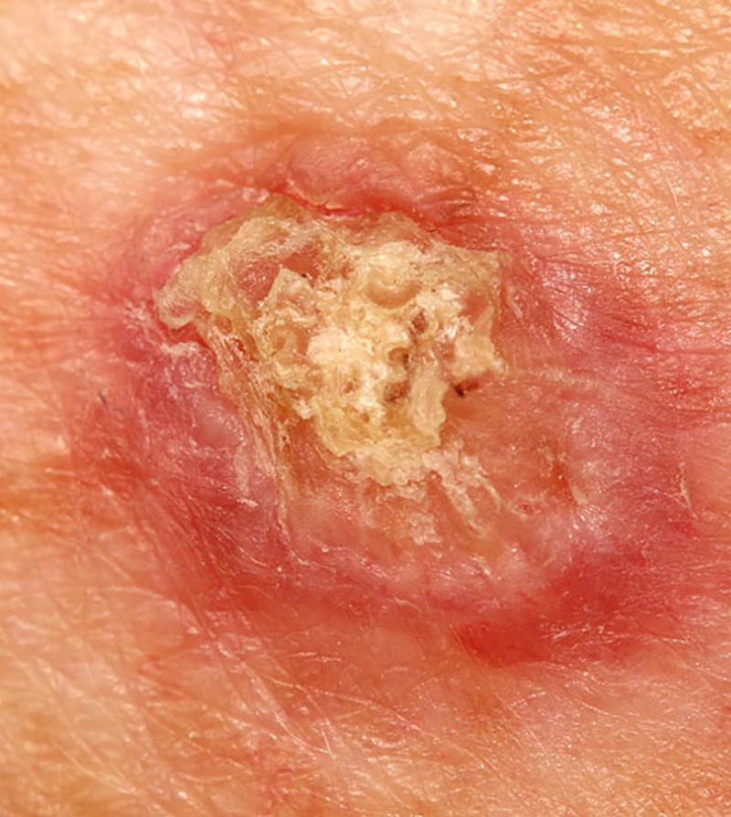
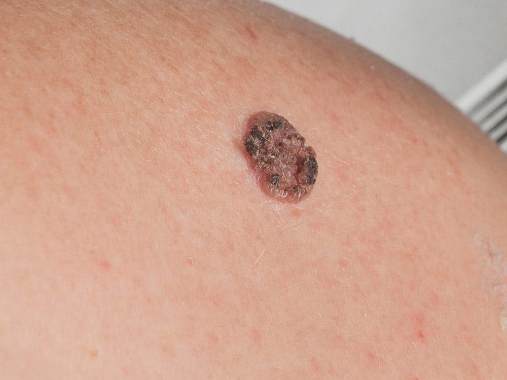
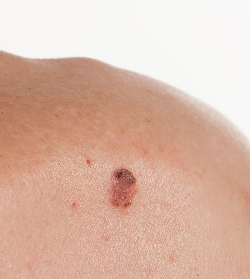
What does squamous cell carcinoma look like?
Squamous cell carcinoma have the following characteristics:
- Crusted-over patch of skin
- Crusted or scaly area of skin with a red inflamed base that resembles a growing tumor
- Non-healing ulcer
What causes squamous cell carcinoma?
Squamous cell carcinoma is caused by excessive sun exposure and patient specific characteristics, such as age, skin type and ethnicity. Other risk factors include immunosuppression, arsenic and ionizing radiation exposure and chronic inflammation.
What are my treatment options?
Squamous cell carcinoma (SCC) can be treated through various methods, depending on the specific circumstances. In cases where the lesion is small and detected early, freezing (cryotherapy) or burning (electrodessication) techniques can be employed to remove the lesions with minimal scarring. Additionally, prescription creams like imiquimod (Aldara) and 5-FU are viable options for treating the lesion.
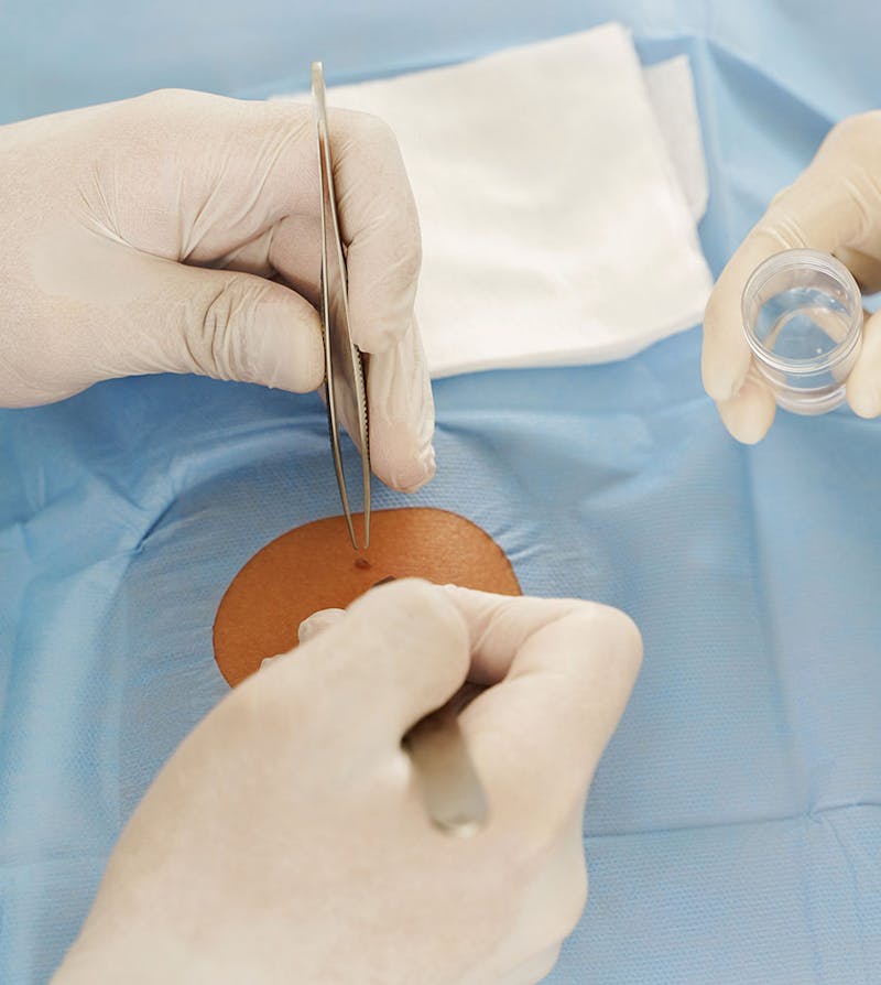
In situations where the lesion is larger or located in a highly visible area, complete excision of the lesion is often recommended to attain optimal aesthetic outcomes.
What happens after the lesion is excised?
Once the lesion is surgically removed and the wound is closed using either permanent or dissolving sutures, it is forwarded to a pathologist for microscopic examination. This step is crucial to confirm the diagnosis and ensure that the entire lesion has been successfully excised.
Following the surgery, a follow-up appointment will be scheduled at the office/clinic after approximately 5-7 days for facial lesions or 10-14 days for lesions located on other parts of the body. During this visit, you will review the pathology report, assess the condition of your incision, and have any necessary sutures removed or trimmed.

What can I do to prevent squamous cell carcinoma?
For individuals who have undergone skin cancer removal and possess multiple risk factors for squamous cell carcinoma (SCC) development, it is recommended to arrange annual skin examinations. These examinations are essential for early detection of any potential lesions, enabling prompt treatment.
Protecting the skin from further sun damage is equally important. Wearing hats and clothing that shield exposed skin, along with the regular application of sunblock, are vital measures to prevent the occurrence of additional skin cancers.